Atomic Force Microscope (AFM)
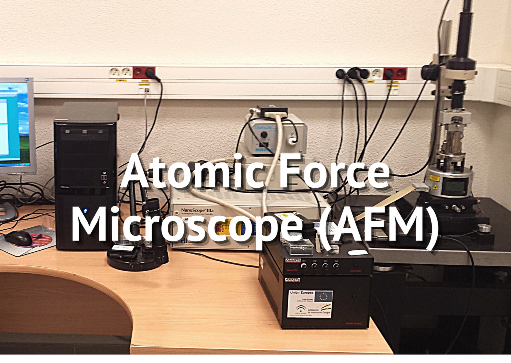
Description of the infrastructure:
Atomic Force Microscope (AFM)
Services currently offered and possible applications in other fields:
Contact mode and intermittent contact topography for 3D imaging of surfaces using atomic force microscopy (AFM), magnetic force microscopy (MFM), and thermal scanning microscopy (SThM), with nanomtric resolution (up to um in vertical resolution and up to 100 um x 100 um in terms of scan size). Determination of surface roughness in this dimensional range. Determination of the size and shape of nano-sized structures, with a lower limit determined by the size of the probe used (minimum 2 nm). Determination and location of magnetic nanometric objects located on a surface of a non-magnetic material, using MFM. Determination of size and shape of regions of different thermal conductivity on a low rough surface, based on SThM. In addition, these techniques can be used to study biological systems (cells, cellular structures, macromolecules, etc) as long as the entities can be deposited on flat support (glass, mica. etc.) and that their size does not exceed team boundaries.
*Rates exclude the price of AFM tips. The user may provide their own tips, as long as they are compatible with the microscope and the technique or mode of work required. Alternatively, the tips necessary to carry out the experiments may be purchased at the SPI itself (subject to availability). In this case, the tips would remain in the possession of the user, upon payment of their cost. As a guide, some tip prices offered by the microscope manufacturer (Bruker) for different working modes are indicated below:
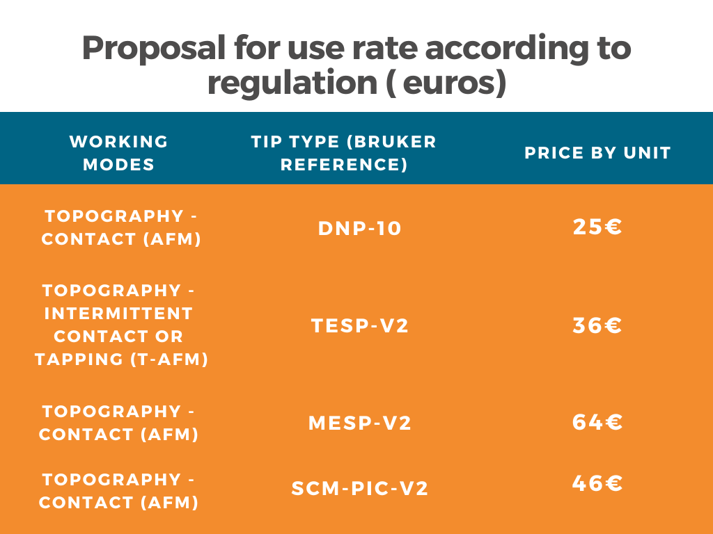
For other types of tips with improved performance or for other advanced working modes, the acquisition prices of the probes should be previously consulted with the technician or scientific manager of the peripheral service.
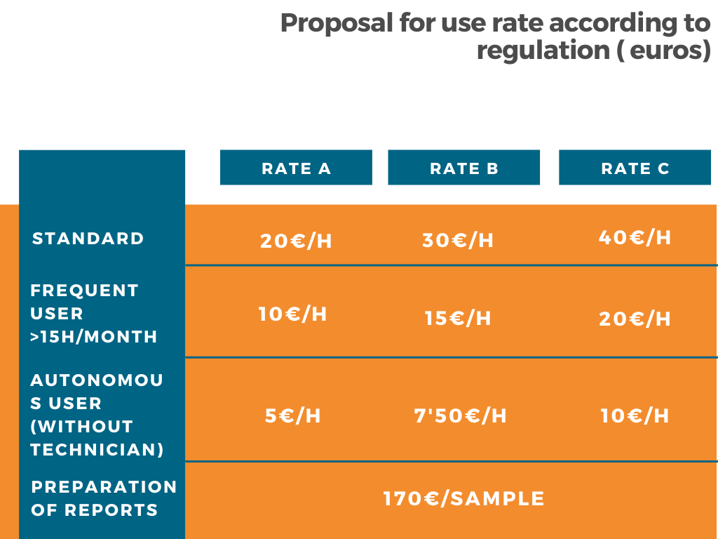
Is it necessary to use a technician: no. Have a technician: no
Self-employed users will be considered those who have successfully completed the training course offered by the SPI AFM/STM. This training course costs €100.
Observations
It is considered that an average work session usually lasts approximately 3 hours. The final result of a ssesion will always be an image or set of images of the sample studied. The technical report, which can be requested additionally, will include the team managers’s interpretation of the images obtained during the session, providing data such as roughness, the size of the structures present, etc.